Global Autism Research & Innovation Summit
THEME: "Connecting Insights, Transforming Lives: A Global Vision for Autism Innovation"
 22-23 Mar 2027
  Paris, France
 THEME: "Connecting Insights, Transforming Lives: A Global Vision for Autism Innovation"
22-23 Mar 2027
Paris, France 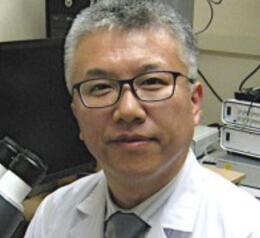
Drexel University College of Medicine, USA
Title: PEDF peptides rescue defects in neurite morphogenesis and intracellular calcium response in cortical neurons from autism mouse model
Kazuhito Toyooka is an Associate Professor in the Department of Neurobiology and Anatomy at Drexel University College of Medicine in Philadelphia, Pennsylvania. He has directed his own research laboratory since 2013. His laboratory studies how the cerebral cortex forms during embryonic development. The laboratory examines neurogenesis, neuronal migration, neurite initiation, neurite formation, synapse formation, and neural connectivity. He also studies the molecular causes of ASD in activity-dependent neuroprotective protein (ADNP) syndrome, and in the neurodevelopmental disorders associated with chromosome 17p13.3. His group combines mouse genetics, primary neuronal culture, in utero electroporation, time-lapse imaging, CRISPR/Cas9 genome editing, RNA sequencing, proteomics, and behavioral analysis. A central goal of the laboratory is the development of intervention, such as peptide therapy and gene therapy, for neurodevelopmental disorders. His research program is supported by the National Institutes of Health and by the Department of Defense.
Autism spectrum disorder (ASD) is a neurodevelopmental disorder. Approved drugs treat irritability in patients with ASD, but the other core symptoms remain untargeted. Pigment epithelium-derived factor (PEDF) is a secreted glycoprotein with neuroprotective functions. Our laboratory previously reported that PEDF regulates neurite formation during cortical development in the mouse. Therefore, we asked whether synthetic PEDF peptides correct neuronal abnormalities in a mouse model of ASD.
We injected valproic acid (VPA) into pregnant mice at embryonic day 9.5 (E9.5). At E14.5, we cultured cortical neurons from control embryos and from VPA-exposed embryos. We added the 44-mer, the 34mer, or the 18-mer PEDF peptide to the culture medium. We measured neurite length and neurite number at 2 days in vitro (DIV2). We measured the density of dendritic spines and the head width of mushroom spines at DIV16. We also recorded spontaneous calcium responses in these neurons with the calcium indicator GCaMP6s.
The longest neurite was shorter in neurons from VPA-exposed mice than in neurons from control mice. The 44-mer and the 18-mer peptides restored the length of the longest neurite. All three peptides restored the reduced number of neurites. The density of dendritic spines was lower in the VPA-exposed neurons than in the control neurons. The head width of mushroom spines was larger in the VPA-exposed neurons than in the control neurons. The 44-mer peptide restored the spine density and the head width to the control levels. Calcium imaging revealed a larger amplitude of the fluorescence change in the VPA-exposed neurons than in the control neurons. The 44-mer peptide reduced this amplitude to the control level.
PEDF peptides correct defects in neurite formation, spine formation, and neuronal activity in this ASD mouse model. Therefore, there is a benefit to further study the efficacy of PEDF peptides as candidate treatments for neurodevelopmental disorders.
